There have been various classifications of acne scars, with often confusing and descriptive nomenclature.
The incidence and severity of various scars in the general population has not been well studied. Acne scars can be broadly divided into three categories:
1. Macular.
2. Atrophic.
3. Hypertrophic.
The atrophic scars are the most common followed by the macular, the least common being hypertrophic scars.
 Macular Scars:
Though these lesions are macules and not strictly scars, they are so-called because they tend to persist for months together.
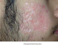
They can be further subdivided into erythematous and hyperpigmented macules. Hypopigmented macules following acne are rare. The hypopigmented lesions seen are true atrophic acne scars rather than macules.
 Erythematous:
Erythematous macules are seen following resolution of inflammatory acne lesions. They are more common in lighter skins and tend to persist in patients that have sensitive skins, photosensitivity or rosacea like skin. They give the impression of active acne lesions and are prone to permanent scarring.
 Hyperpigmented:
Hyperpigmented macules occur due to postinflammatory hyperpigmentation. They can be very persistent and are more common in darker skinned patients and in those patients who constantly pick at their lesions (Acne excoriee).
Depressed Scars:
Depressed atrophic scars are the most common type of scars
seen following acne. They can be further subdivided according to the shape, depth and size.
 Ice Pick Scars:
These are one of the most common scars seen in acne and also the most difficult to treat. Ice pick scars are deep, narrow and sharply defined epithelial tracts that extend vertically to the deep dermis or subcutaneous tissue. The surface opening is wider than the deeper portion as it tapers into a ‘V’ shape. They appear as if the skin has been pricked with an ice pick, which is an instrument with a tapering sharp point.
They tend to worsen with age as the skin becomes loose and appear as open pores giving a mature look to the patient’s skin. These scars are more commonly seen on the cheeks, glabellar region and nose.
 Boxcar Scars:
Boxcar scars are round, oval or irregular sharply defined scars with vertical edges, similar to varicella scars. They are called boxcar scars because on cross-section they appear sharply punched out like a boxcar which is a wagon of a goods train.
They are wider at the surface than ice pick scars, appear punched out and do not taper to a point at the base. They may be shallow (0.1–0.5 mm) or deep (>0.5 mm) and are most often 1.5 to 4.0 mm in diameter. They are most common on the temples and cheeks.
 Rolling Scars:
Rolling scars are wide undulating scars, with relatively normalappearing overlying skin.
They occur due to dermal tethering of the epidermis and dermis to the underlying subcutis leading to superficial shadowing and a rolling or undulating appearance. They appear most common on the lower cheeks and mandibular area and worsen with age.
 Linear Scars:
These scars have not been commonly described in reported literature, though they are often observed. They appear as linear atrophic lines that are commonly hypopigmented with relatively normal skin in between. They may be narrow linear scars, where they appear as thin lines connecting each other or broad linear scars, appearing as wider linear dermal depressions.
 Lipoatrophic Scars:
These scars occur following resolution of cystic acne, which destroys the fat. They are more common in males with a thin asthenic face, giving the face a gaunt appearance.
 Elevated Scars
Elevated scars are less common following acne. They may be of different types.
Hypertrophic Scars
They are elevated, dome-shaped fibrotic scars, skin colored or hyperpigmented, frequently seen in the mandibular area of the face and back.
They are more common in males and in darker skin types. They remain localized and do not extend beyond the surface of the acne lesions.
 Keloidal Scars
These are keloids developing in acne lesions, which extend beyond the original acne lesions. They have irregular borders and are often symptomatic causing itching or pain. They are seen more often in males, on the back, shoulders and chest.
 Papular Scars
These scars appear as raised, hypopigmented and papular lesions, most commonly seen on the back, chin and nose (Figs 2.12Aand B). They occur due to destruction of the perifollicular dermis and are in fact outpouchings of the epidermis due to loss of support of the underlying dermis.
 Bridging Scars and Sinus Tracts
These are channels joined together by epithelial tracts overlying normal or atrophic skin. They often contain foul smelling products of sebum and cause great distress.